IMAGES TAKEN FOR STANFORD MEDICINE’S LAB OF MARIA BARNA FOR A STORY ABOUT RIBOSOMES:
Behind the Science: Ribosomes, the protein factories of cells
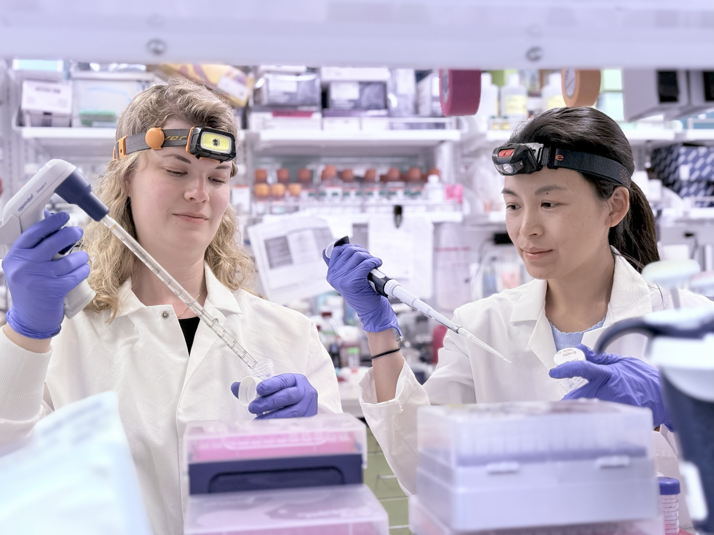
1. SEPARATION: Researchers make different solutions that help break open cells, clean up the contents inside those cells, and separate out the ribosomes.

2. ISOLATION: The researchers then pipette a solution to isolate ribosomes in live cells. This solution has special "tags" and a molecule that stops the ribosomes in place. The team works in the dark with red lights to prevent triggering a light-based reaction too early. A biosafety cabinet maintains a sterile environment.

3. MODIFICATION: The cells have now been modified so that they can tag ribosomes when exposed to the blue light of the incubator. The incubator is kept at 37 degrees Celsius, which is the ideal temperature for mouse embryonic stem cells to survive and grow.
4. IDENTIFICATION: The researchers use a powerful microscope to view all the tagged ribosomes in the cell at once, which helps them identify different groups of ribosomes and how they work together. A single mammal cell can have up to 10 million ribosomes.

5. CLASSIFICATION: In fluorescently tagged ribosomes, the different colors show different parts of the ribosome: Green represents the large subunit, red represents the small subunit and yellow indicates where both parts come together to form a complete ribosome.